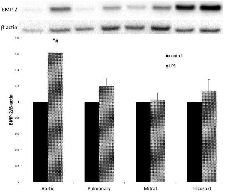
Figure 4.

Aortic VICs adopt an osteogenic phenotype after TLR stimulation, while the pulmonic, mitral, and tricuspid VICs do not. VICs were lysed after stimulation with LPS (200ng/mL) for 48 hours. Lysates were analyzed using immunoblotting. Levels of BMP-2 increased significantly (*p<0.05) compared to control in the aortic valve, but not the other three heart valves after LPS stimulation. Also, BMP-2 levels after LPS stimulation in Aortic VICs were greater compared to LPS-stimulated BMP-2 levels in the other three heart valves (#p<0.05). Data are normalized to control for each valve.
