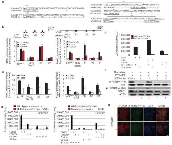
Figure 5.
FOXO1 activates the expression of OCT4 and SOX2 pluripotency genes in hESCs by binding directly to their regulatory regions. (a) Sequence alignment of human OCT4 and SOX2 regulatory regions containing putative FoxO-binding sites. (b) Endogenous FOXO occupation of sites shown within arrows was analysed by ChIP in H1 cells. DNA co-immunoprecipitated with either anti-FOXO1 (H-128), anti-FOXO3A (N-15) or a control pre-immune immunoglobulin was amplified by qPCR. Binding of FOXO1 to p27KIP1 promoter and binding of FOXO1 to a conserved upstream region with no FOXO-binding sequences (in 2.7 kb of human OCT4 (NEG Seq O) and in -4 kb of human SOX2 (NEG - Seq S)) were used, respectively, as positive and negative controls. Results of three independent experiments (mean ± s.e.m.) are shown as relative fold enrichment when compared with control immunoglobulin after normalization to the values obtained from the input samples. (c) ChIP analysis of FOXO1 in undifferentiated H1/FOXO1-shRNA hESCs treated or not with doxycycline (Dox) for 3 days. Results of three independent experiments (mean ± s.e.m.) are shown. (d) pcDNA3, pcDNA3-FOXO1 or pcDNA-FOXO3A were co-transfected into HEK293T cells with a pGL3 containing the O1 or O2 site of human OCT4, or the S1, S2 or S3 site of SOX2 or their mutants driving the luciferase gene; luciferase activity was measured 48 h later. Results are from three independent experiments (mean ± s.e.m.). (e) Luciferase activity measured 48 h after co-transfection of hESCs (H1) with a lentiviral vector control or containing FOXO1 or FOXO3A and a human OCT4 reporter plasmid containing a wild-type or mutant O2 sequence. Results are the mean of two independent experiments. (f) H1 hESCs were maintained continuously (Contin.) in bFGF for 72 h (lane 1) or starved overnight and stimulated or not with bFGF (40 ng ml−1 for 10 min) and/or PI(3)K inhibitor LY294002 (10 μM) for 45 min before stimulation, and preparing the whole-cell lysate (lanes 2-5). One representative immunoblot of two experiments is shown. (g) H1 cells maintained in undifferentiated conditions and treated or not with LY294002 (10 μM) for 2 h before double immunostaining of FOXO1 and phospho-AKT (Ser 473) and counterstaining with DAPI. One representative of three independent experiments is shown (uncropped scanned gels are shown in Supplementary Fig. S12).

