Fig. 4.
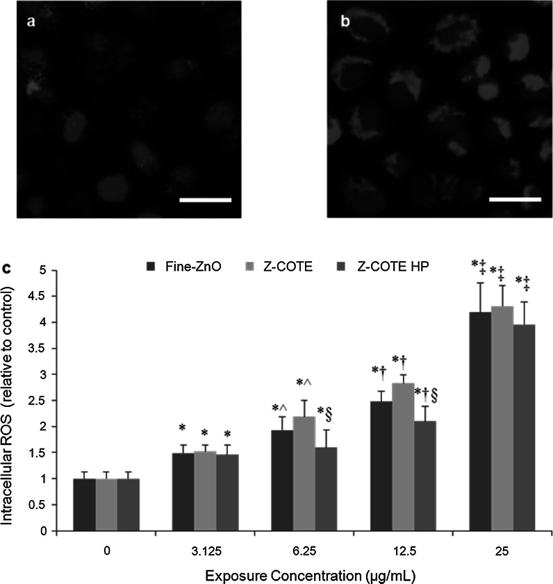
The effect of ZnO-NPs on cellular oxidative stress. a, b The expression of reactive oxygen species (ROS) in living FE1-MML cells after exposure to even a low concentration of Z-COTE (6.25 µg/mL). a non-treated cells; b Z-COTE-treated cells. Intracellular ROS was identified by a fluorogenic probe (red, cytoplasm) and DAPI (blue, nucleus). Scale bar = 25 µm. c ROS data demonstrated that ZnO-NPs elevated intracellular ROS in a dose-dependent manner (*p < 0.05 vs. control cells; ^ p < 0.05 vs. 3.125 µg/mL; † p < 0.05 vs. 6.25 µg/mL; ‡ p < 0.01 vs. 12.5 µg/mL; n = 6), and Z-COTE HP1 (surface coating with triethoxycaprylysilane) showed a lower level (§ p < 0.05, n = 6) of ROS compared to Z-COTE (uncoated ZnO-NPs). (Color figure online)

