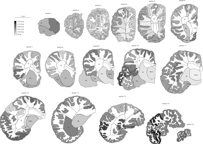
Figure 4.
Distribution of neuronal densities along the cerebral cortex of the African elephant. Coronal sections, 1.28 mm thick, are numbered from the anterior to the posterior pole. Color intensity of the delineated cortical grey matter indicates the local neuronal density according to the scale on the left. A-I, blocks of cortical grey matter processed separately. W, white matter. Call, corpus callosum, processed separately from the remaining white matter. Amyg, amygdala; Dienc, diencephalon; Ent, entorhinal cortex; Hp, hippocampus; Str, striatum.

