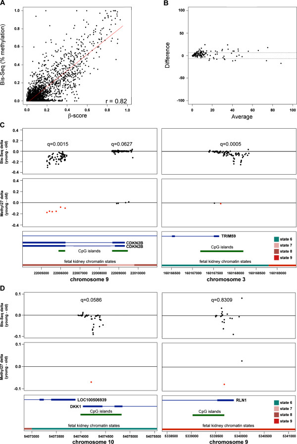
Figure 5.

Validation of ageCGs by targeted capture and bisulfite sequencing of genomic regions encompassing ageCG sites. (A) Scatterplots of percentage methylation by bisulfite sequencing (Bis-seq) versus Methylation27 β-score and correlation of methylation values between the two methods across 19 kidney samples used for validation. (B) A representative Bland-Altman plot for comparison of Bis-seq and Methylation27 methylation values. Points depict the average percentage methylation between both methods plotted against the differences in methylation between the methods. Dotted lines show limits of agreement (average difference ± 1.96 standard deviation of the difference). (C, D) Comparison of methylation delta values (median percentage methylation of young minus old samples) at CpGs covered by Bis-seq (top panels) and Methylation27 arrays (middle panels). Red points represent ageCGs, and delta values shown for Bis-seq are only for the 9 youngest and 10 oldest samples. False discovery rate (q) values indicate significance level of a widespread age effect by linear mixed model results with Bis-seq data at these target regions. The DKK1 target just missed the significance threshold (q < 0.05), and the RLN1 target demonstrates an example region that contained abrupt changes in direction of delta values among neighboring CpGs. Bottom panels depict labeled gene regions (blue boxes, exons blue lines, introns), CpG islands (green boxes), and fetal kidney chromatin states (colors correspond with definitions in Figure 4). Greater details and examples of all Bis-seq targets are available in Additional files 7 and 24.
