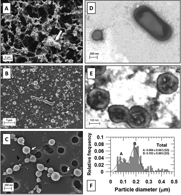
FIG 2.
The two cell size populations and nanoparticles of Lake Vida brine. (A) SEM of fixed brine prepared on a 0.2-μm-pore-size polycarbonate filter, air-dried (not dehydrated). A microbial cell trapped on the filamentous network is indicated by the white arrow. (B) SEM of fixed and dehydrated brine prepared on a 0.2-μm-pore-size polycarbonate filter showing the brine cells surrounded by a dense layer of organic and inorganic material covering the filter (volume of brine filtered, 1 ml). (C) SEM of fixed and dehydrated Lake Vida ultrasmall microbial cells prepared on a 0.2-μm-pore-size polycarbonate filter (volume of brine filtered, 0.5 ml). Nanoparticles attached to the cell surface (indicated by black arrows) and uncharacterized filaments connecting the cells (indicated by white arrows). (D) STEM of fixed and dehydrated brine cells treated with EDTA and stained with UA, pH 4 to 5. A cell of ∼0.2 μm in diameter is shown on the left of the image, and a rod-shaped bacterial cell is shown on the right. (E) STEM of ultrasmall brine cells stained with PTA, pH 0.4. Scale bars are shown in each micrograph. (F) Size distribution of cells and unidentified nanoparticles in Lake Vida brine.

