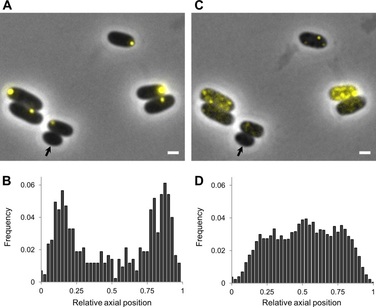
FIG 1.
In vivo HP exposure leads to PA disassembly in E. coli MG1655 ibpA-yfp. (A and C) Microscopic images show the same MG1655 ibpA-yfp cells before (A) and after (C) HP exposure (300 MPa, 20°C, 15 min). Phase-contrast images are superimposed with YFP epifluorescence images (reporting PAs), and the scale bar corresponds to 1 μm. The black arrow indicates a cell devoid of PAs. (B and D) Binned histograms show PA distribution along the relative axial position of the cells as detected in untreated (B) and HP-exposed (300 MPa, 20°C, 15 min) (D) MG1655 ibpA-yfp cells. The average numbers of PA foci per cell were 1.00 for control cells (n = 427) and 5.45 for HP-exposed cells (n = 640).

