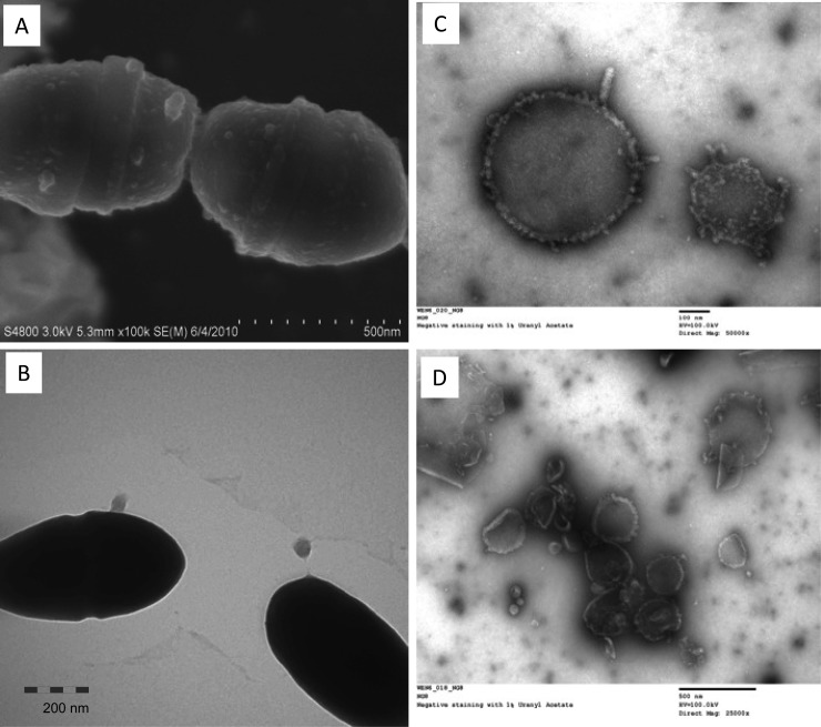
FIG 5.
EM analysis of S. mutans membrane vesicles. S. mutans UA159 (A) and NG8 (B to D) were grown in BHI broth (B to D) and in biofilm medium on hydroxylapatite discs (A) overnight. FE-SEM (A) and TEM (B) analysis show small blebs on the cell surfaces. Panels C and D show EM images of vesicular structures in cell-free supernatants of NG8 following negative staining with 1% uranyl acetate. Similar vesicles were also seen with UA159 (not shown). Images were taken at magnifications of 50,000× (A), 30,000× (B), 50,000× (C), and 25,000× (D).

