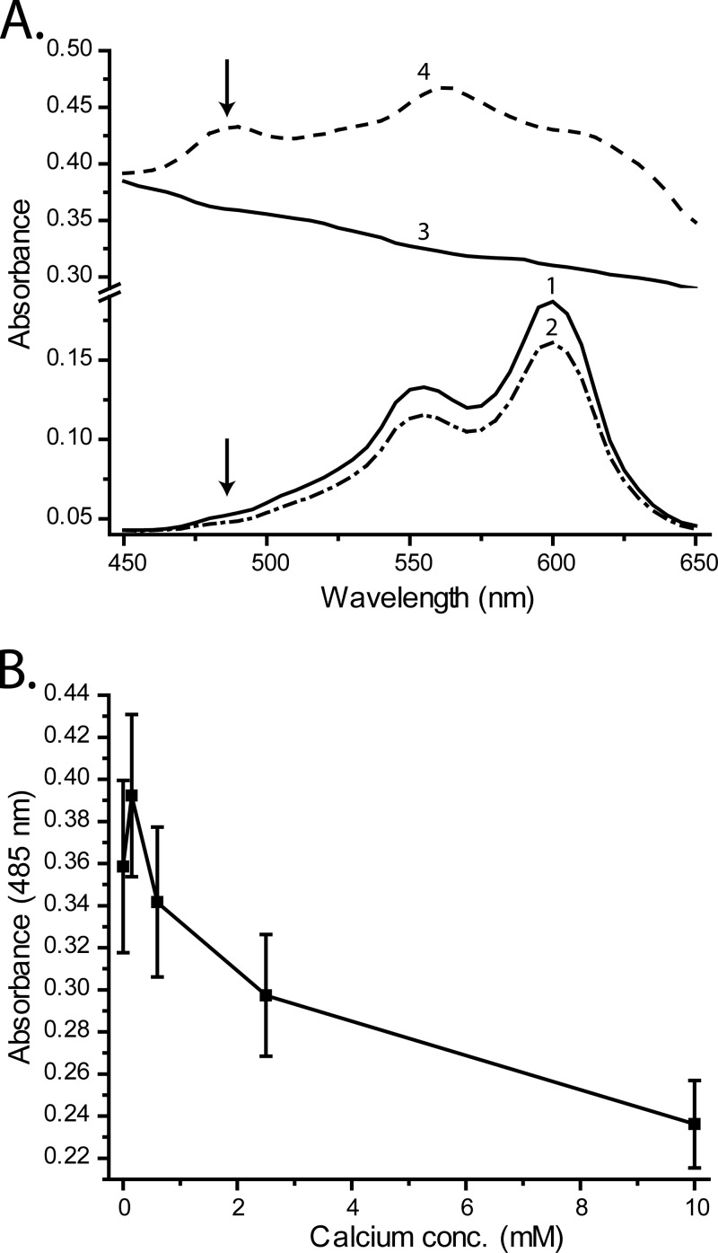
FIG 8.
Pinacyanol dye binding to RP1137 cells is disrupted by the addition of calcium. (A) Absorbance spectra of water plus dye (curve 1), water plus dye plus 10 mM CaCl2 (curve 2), RP1137 cells alone (curve 3), and RP1137 cells plus dye (curve 4). Arrows indicate the position of the 485-nm absorbance band indicative of pinacyanol binding of teichoic acid. (B) Absorbance of pinacyanol-stained cells at 485 nm with increasing calcium concentration. Data are means and standard errors for three biological replicates with two technical replicates each.

