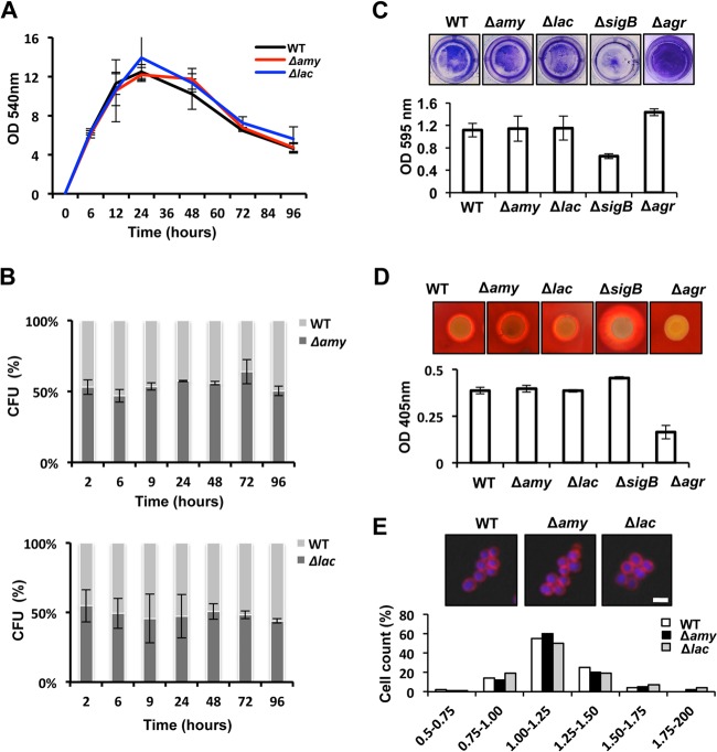
FIG 2.
Physiological assays of the wild-type strain and the amy- and lac-defective mutants. (A) Growth analysis of wild-type (WT) strain Newman and the Δamy and Δlac strains. The OD540 was monitored for 96 h in TSB cultures grown at 37°C. (B) Competition assays of mixed populations (1:1 ratio) of WT and Δamy::YFP strains (Δamy) and of WT and Δlac::YFP strains (Δlac). Mixed communities were incubated at an initial OD540 of 0.05. Samples were taken over 96 h, and CFU were counted by using a dissection scope equipped with a fluorescence excitation detection system. (C) Biofilm formation assay of cultures grown in 24-well titer plates. Biofilms were stained with crystal violet (1%) and quantified by spectrophotometry analysis (n = 3) (error bars represent standard deviations). (D) Hemolytic activity of the different strains. Cultures grown overnight were spotted onto TSB agar containing 5% sheep blood and incubated at 37°C during 48 h. Quantification of hemolytic activity was performed by measuring the OD405 of a solution of 2% erythrocytes previously incubated with the strains' supernatants (n = 3) (error bars represent standard deviations). (E) Fluorescence microscopy pictures used to monitor the cell sizes of the different strains. Cultures were grown until they reached the exponential phase. The membrane of the cell was stained with Nile red (false colored in red), and the DNA was stained with Hoechst 3342 (false colored in blue). Quantification of the cell diameter was performed with Leica Application software (see Materials and Methods).

