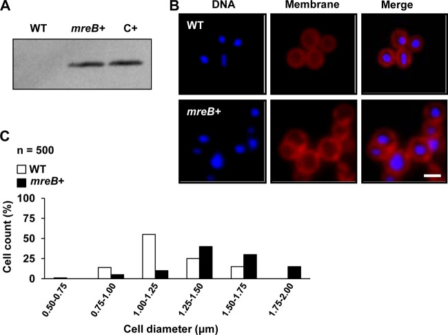
FIG 3.
S. aureus cells expressing mreB of B. subtilis. (A) Western blot analysis detecting the presence of the MreB protein in cell extracts by using polyclonal antibodies against MreB of B. subtilis. WT is wild-type strain Newman. mreB+ is a Newman strain expressing mreB of B. subtilis under the control of an IPTG-inducible promoter. The positive control (C+) is B. subtilis strain PY79. (B) Comparison of cell sizes of wild-type and mreB+ strains. Cultures were grown until exponential phase was reached, and the cell size was monitored by fluorescence microscopy. The cellular membrane was stained with Nile red (false colored red), and the DNA was stained with Hoechst 3342 (false colored blue). Bar, 1 μm. (C) Quantification of the diameters of 500 cells selected from wild-type and mreB+ microscopic fields.

