
FIG 6.
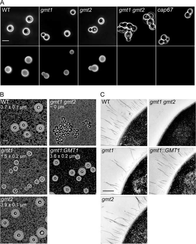
Capsule alteration in gmt mutants. (A) Antibody staining. Bright-field images of the indicated strains grown under capsule-inducing conditions and labeled with anticapsule antibody 3C2 (see Materials and Methods) (top); immunofluorescence micrographs of the same fields (bottom). Scale bar, 5 μm. (B) Negative staining. The indicated strains were grown as indicated for panel A, mixed with India ink as described in Materials and Methods, and examined by light microscopy. The average and standard deviation of the mean capsule thickness for 25 cells is indicated on each panel; this could not be assessed for the apparently acapsular and highly clumpy double mutant. Scale bar, 5 μm. (C) Electron microscopy. Each image shows part of the edge of one representative cell for the strain indicated. gmt1::GMT1 denotes the gmt1 mutant chromosomally complemented with the wild-type gene. Scale bar, 250 nm.