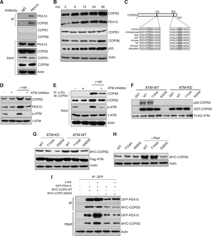
FIG 2.
PEA15 protein stability is regulated by the proteasomal degradation pathway. (A) Immunoblot analysis for indicated proteins after immunoprecipitation (IP) with IgG control or PEA15 antibody. Input was also analyzed for the indicated proteins. (B) Immunoblot analysis of the indicated proteins at the indicated time points after gamma irradiation. Actin was used as a loading control. (C) Schematic indicating two evolutionarily conserved potential ATM kinase phosphorylation sites. (D) Immunoblot analysis for the indicated proteins before and after gamma irradiation with or without treatment with the ATM inhibitor (10 μM). (E) Immunoblot analysis of COPS5 after immunoprecipitation of the phospho-SQ fraction of HCT116 cells isolated under the indicated conditions. As controls, the indicated proteins were analyzed in the input. (F) Autoradiograph of 32P-labeled GST–COPS5-WT, GST–COPS5-T154A, and GST–COPS5-S320A after incubation with ATM or ATM-KD. Immunoblots of GST-COPS5 substrates and FLAG-ATM are also shown. (G) Immunoblot analysis of the indicated proteins in HCT116 cells expressing COPS5 WT, T154A, and S320A proteins along with ATM-FLAG KD or ATM-FLAG WT plasmids. (H) Immunoblot analysis of MYC-COPS5 and actin in HCT116 cells ectopically expressing COPS5 WT, T154A, or S320A proteins left untreated or irradiated with 20-Gy gamma radiation. (I) Immunoblot analysis after immunoprecipitation (IP) with PEA15 antibody in HCT116 cells that were unirradiated or gamma irradiated. The indicated proteins were analyzed in the inputs as controls.

