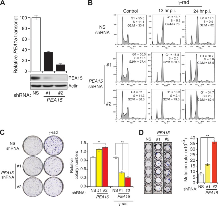
FIG 5.
Loss of PEA15 causes DNA damage-induced G2/M checkpoint defect and increased mutagenesis. (A) RT-qPCR analysis of PEA15 mRNA levels (n = 3) (top) and immunoblot analysis of PEA15 levels (bottom) in HCT116 cells expressing NS or PEA15 shRNAs (1 and 2). Actin was used as a loading control. Error bars indicate standard errors of the means. (B) Flow cytometry analysis to determine the cell cycle distribution of unirradiated (control) or gamma-irradiated HCT116 cells expressing the indicated shRNAs. Gamma-irradiated cells were collected at 12 or 24 h after irradiation. The percentage of cells in each stage of the cell cycle is indicated. (C) Clonogenic assay to monitor the survival of unirradiated and gamma irradiated (γ-rad) HCT116 cells expressing the indicated shRNAs (n = 3). Quantification of relative colony numbers is presented. Error bars indicate standard errors of the means. *, P < 0.01; **, P < 0.001. (D) Spontaneous mutation of the hprt gene (n = 3). Quantification of the mutation rate under the indicated conditions. Error bars indicate standard errors of the means. **, P < 0.001. Representative wells of crystal violet stained HCT116 cells expressing indicated shRNA that were grown in 6-TG are shown on the left.

