FIG 6.
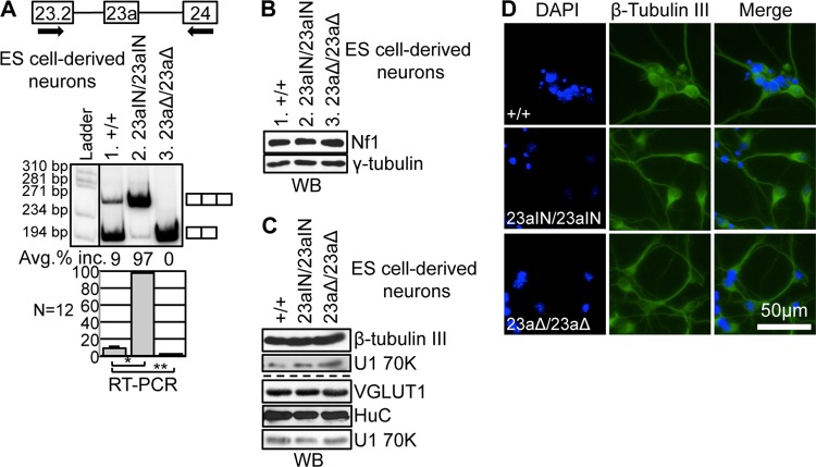
Manipulation of Nf1 exon 23a inclusion in ES cell-derived neurons. (A) RT-PCR showing endogenous Nf1 exon 23a inclusion in ES cell-derived neurons. Arrows indicate locations of RT-PCR primers. The RT-PCR band size was 266 bp if exon 23a was included and 203 bp if exon 23a was skipped. Error bars represent standard errors. *, P = 1.5 × 10⁻²⁴; **, P = 1.5 × 10⁻⁴. (B) Western blot analysis showing Nf1 protein (∼250 kDa) expression in ES cell-derived neurons. γ-Tubulin (∼48 kDa), a ubiquitously expressed protein, was used as a loading control. (C) Western blot showing expression of the neuronal marker β-tubulin III (∼55 kDa), the glutamatergic neuron marker VGLUT1 (∼61 kDa), and the neuron-enriched protein HuC (∼39 kDa) in ES cell-derived neurons. U1 70K (∼70 kDa) was used as a loading control. (D) Immunofluorescence images demonstrating that ES cell-derived neurons express the neuronal marker β-tubulin III and show neuron-like morphology.

