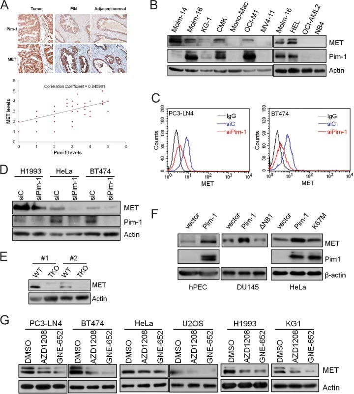
FIG 1.
The Pim-1 kinase regulates MET expression. (A) Representative images of a human prostate tissue microarray stained with anti-MET and anti-Pim-1 antibodies against normal, prostatic intraepithelial neoplasia (PIN), and tumor tissues. The relative strength of antibody staining was plotted as Pim-1 versus MET. The correlation coefficient (R) was derived by Microsoft Excel analysis. (B) Cell lysates from a panel of acute myeloid leukemia cell lines were analyzed by immunoblot assays using the indicated antibodies. (C) PC3-LN4 and BT474 cells were treated with siRNA targeting Pim-1 (siPim-1) or a nontargeting control (siC) for 72 h. The level of cell surface MET expression was visualized by flow cytometry using anti-MET antibody and R-phycoerythrin (PE)-conjugated secondary antibody. (D) Cell lysates from H1993, HeLa, and BT474 cells treated with siRNA targeting Pim-1 (siPim-1) or a nontargeting control (siC) for 72 h were analyzed by immunoblot assays using the indicated antibodies. (E) Cell lysates from wild-type (WT) and Pim TKO mouse embryonic fibroblasts were analyzed by immunoblot assays. Two different pairs of WT and TKO cells (#1 and #2) isolated separately from different embryos are examined. (F) Human prostate epithelial cells (hPEC), DU145 cells, or HeLa cells were transfected with plasmids expressing Pim-1 or its kinase-dead mutant ΔN81 or K67M. After 48 h, the levels of Pim-1, MET, and actin were examined by Western blotting. (G) Various tumor cell lines were treated with Pim inhibitor AZD1208 (3 μM) or GNE-652 (1 μM) for 24 h. Cell lysates were analyzed by immunoblot assays using the indicated antibodies.

