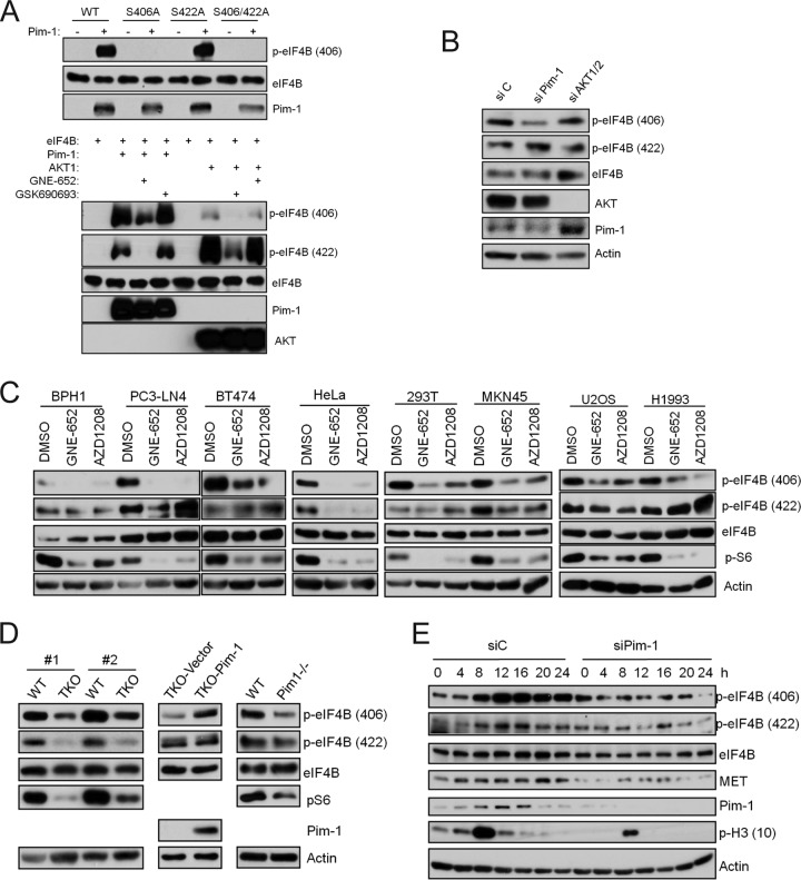
FIG 3.
Pim-1 controls eIF4B phosphorylation at S406. (A) (Top) FLAG-tagged eIF4B as well as its mutants was expressed in U2OS cells and immunoprecipitated using anti-FLAG antibody with protein A/G beads. The beads were extensively washed and incubated with 100 ng of purified Pim-1 proteins at 37°C for 30 min. Samples were analyzed by immunoblot assays using indicated antibodies. (Bottom) Purified eIF4B and Pim-1 or AKT1 were incubated in the presence or absence of GNE-652 (0.1 μM) or GSK690693 (0.1 μM). Samples were subjected to immunoblotting analysis with the indicated antibodies. (B) HeLa cells were transfected with Pim-1 siRNA, AKT1/2 siRNA, or a nontargeting control siRNA for 72 h. Cell lysates were analyzed by immunoblot assays using indicated antibodies. (C) Cell lines were treated with GNE-652 (1 μM) or AZD1208 (3 μM) for 3 h. Cell lysates were analyzed by immunoblot assays using indicated antibodies. (D) Cell lysates from two independently isolated pairs of WT and Pim TKO MEF cells (left), TKO MEFs with empty vector (TKO-Vector) and TKO MEFs with a Pim-1-expressing construct (TKO-Pim-1) (middle), and WT MEFs and Pim-1 single-knockout MEFs (right) were subjected to Western blotting with the indicated antibodies. (E) HeLa cells were synchronized with a double-thymidine block protocol and simultaneously treated with Pim-1 siRNA. Cells were released into fresh medium and harvested at indicated times. Cell lysates were subjected to Western blotting with the indicated antibodies.

