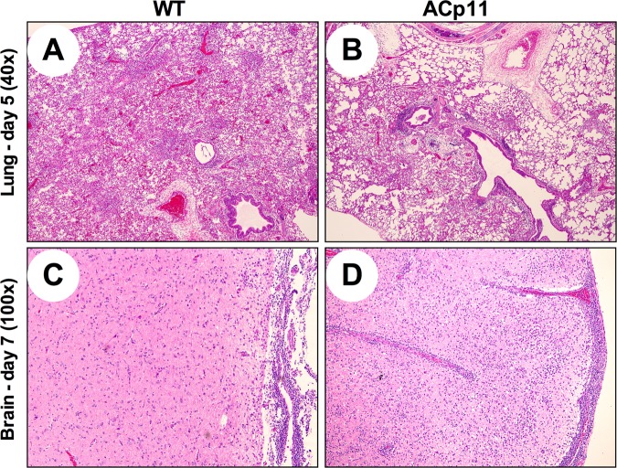
FIG 7.
Histopathology of lung and brain samples showing differences in inflammation between animals infected with WT H7N1 or ACp11 virus on dpi 5 and 7. Ferrets (n = 6/virus) were infected with either WT H7N1 or ACp11. On dpi 3, 5, and 7, lung, trachea, and brain samples were collected for evaluation of histopathology. All sections were fixed and subjected to H&E staining. Sections were viewed and scored by a veterinary pathologist blinded to the virus strain's identity. Shown are representative images of dpi 5 lung sections from WT H7N1 (A)- and ACp11 (B)-infected ferrets and dpi 7 brain sections from WT H7N1 (C)- and ACp11 (D)-infected ferrets.

