
FIG 1.
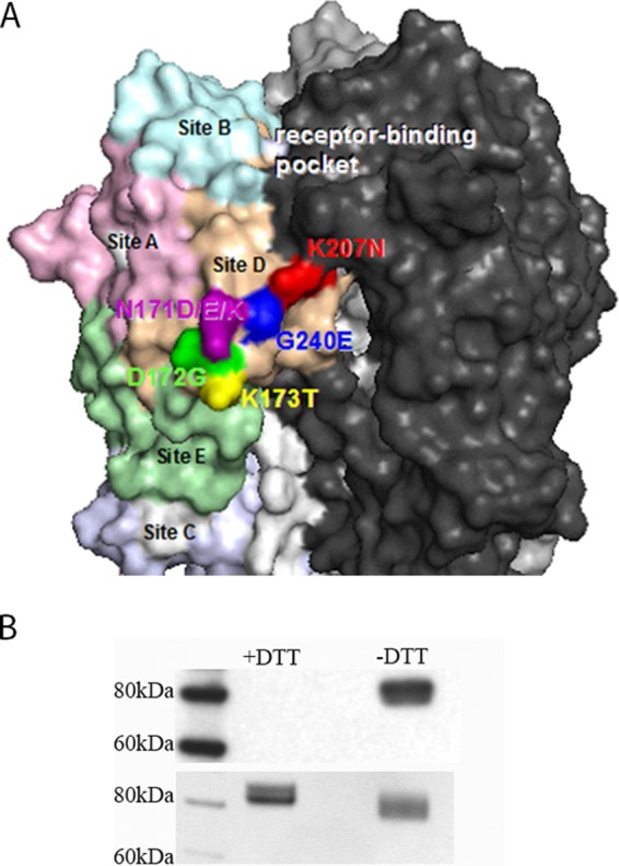
Characteristics of the putative D1-8 antigenic site. (A) D1-8 epitope illustrated on a space-filling model of the trimeric membrane-distal globular head of H3 HA. The H3 HA antigenic sites are colored pink (site A), light blue (site B), light purple (site C), tan (site D), and light green (site E). The D1-8 MAb escape mutations are labeled by amino acid letters and residue numbers. Residues 171 (dark purple), 172 (dark green), 173 (yellow), 207 (red) and 240 (dark blue) are proximal to antigenic site D, which is adjacent to the receptor binding pocket. (B) Western blot analysis of the D1-8/H3 HA interaction. (Top blot) The recombinant trimeric HA of A/Perth/09 proteins were disrupted in 2% SDS without (−) DTT or with (+) 100 mM DTT and then analyzed by Western blotting with MAb D1-8. (Bottom blot) A Coomassie blue-stained duplicate gel to show equal loading of HA proteins.