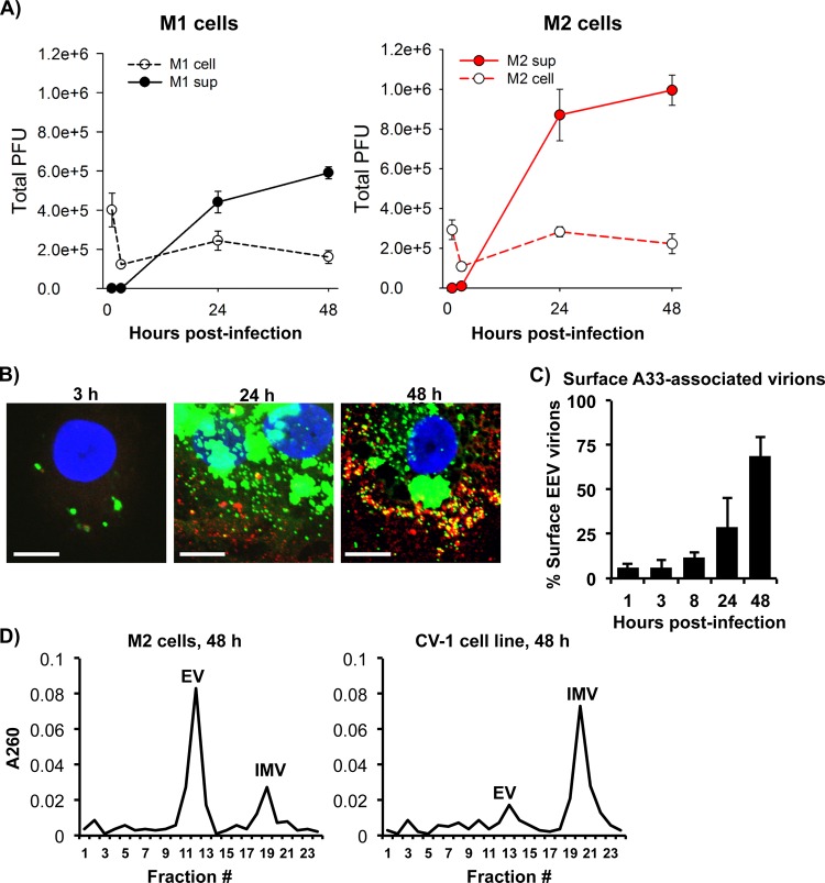
FIG 3.
MDMs mainly produced enveloped forms of VACV. (A) M1- and M2-polarized MDMs were infected with VACV WR at an MOI of 5. Virions were extracted from either infected cells or culture supernatant (sup) at 3 h, 24 h, and 48 h postinfection and subjected to virus titration using the virus plaque assay. (B) M2-polarized cells were infected with vA5L-YFP (green) at an MOI of 5 for various times as indicated and then subjected to surface staining of VACV envelope protein A33 (red) with confocal microscopy analysis. Scale bars represent 10 μM. (C) The number of extracellular (vA5L-YFP plus A33 staining) and intracellular (vA5L-YFP only) cell-associated virions were counted at different time points as indicated. (D) Purified VACV particles from VACV WR-infected M2-polarized cells and CV-1 cells were extracted from cell lysates and supernatants and analyzed by separation on a CsCl density gradient. Fractions from the gradients were tested for absorbance at 260 to estimate the amount of virus particles corresponding to the buoyant densities of mature or enveloped forms of VACV. All data are representative of cells derived from five blood donors. EV, enveloped virus; IMV, intracellular mature virus.

