
FIG 4.
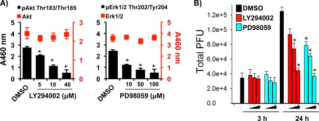
VACV-associated signaling pathways were required for VACV replication in MDMs. M2-polarized MDMs were infected with VACV WR at an MOI of 5 for 3 h. Cells were washed and treated with either 5 μM, 10 μM, or 40 μM LY294002 (Akt inhibitor) or 1 μM, 10 μM, or 100 μM PD98059 (Erk inhibitor). Cell lysates were collected at 3 h, 24 h, and 48 h postinfection. DMSO, dimethylsulfoxide. (A) Lysates were analyzed by a sandwich ELISA coated with anti-Akt or anti-Erk1/2 Abs and treated with secondary Abs against pAkt, pErk, or total target proteins. (B) Virions were extracted from cellular lysates at the time points indicated and titrated by the virus plaque assay. All data are representative of cells derived from five blood donors. *, P < 0.05.