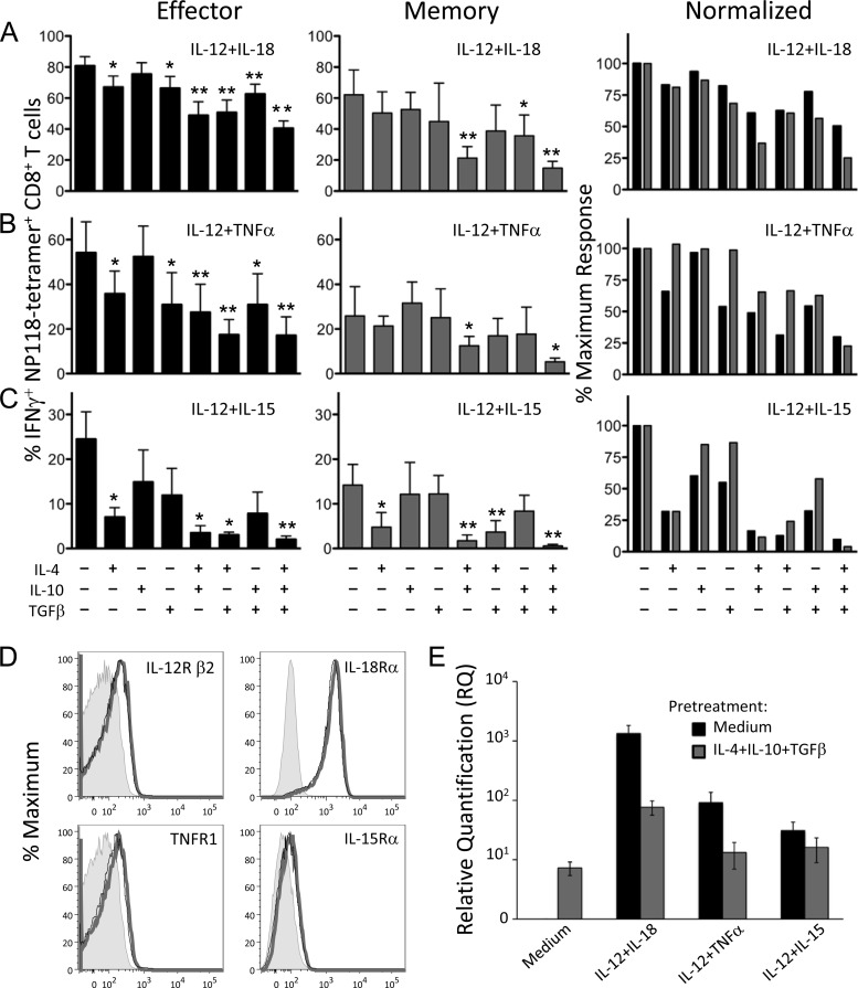
FIG 2.
Exposure to anti-inflammatory cytokines inhibits innate IFN-γ production by CD8+ T cells. Splenocytes from BALB/c mice at 8 days (effector) or >60 days (memory) after LCMV infection were treated directly ex vivo with combinations of IL-4, IL-10, and TGF-β (10 ng/ml each) for 2 h, followed by stimulation with IL-12 plus IL-18 (A), IL-12 plus TNF-α (B), or IL-12 plus IL-15 (C) at 10 ng/ml for 6 h. Brefeldin A was added to cultures for the final hour of incubation, and cytokine production was assessed by intracellular cytokine staining and flow cytometry. Numbers represent the percentage of NP118 tetramer+ CD8+ T cells producing IFN-γ and are the average ± SD from 4 to 6 mice. IFN-γ responses of T cells pretreated with inhibitory cytokines were compared to those of cells pretreated with medium alone using an unpaired two-tailed Student's t test. Inhibitory cytokine combinations that significantly reduced IFN-γ production are marked with asterisks: *, P < 0.05; **, P < 0.01. For normalization, IFN-γ responses from day 8 (black bars) or immune responses (gray bars) are graphed as a percentage of the maximum response to proinflammatory cytokine stimulation with no IL-4, IL-10, or TGF-β added. (D) Cytokine receptor expression on virus-specific CD8+ T cells. Splenocytes from BALB/c mice at 8 days post-LCMV infection were treated with IL-4 plus IL-10 plus TGF-β or medium alone for 2 h at 37°C. Levels of IL-12Rβ2, IL-18Rα, TNFR1, and IL-15Rα on NP118 tetramer+ CD8+ T cells were examined by flow cytometry. Gray-shaded histograms represent unstained controls. Compared to cells treated with medium alone (thick gray line), cells treated with IL-4 plus IL-10 plus TGF-β (thin black line) showed no significant difference in expression levels of IL-12Rβ2 (P = 0.48), IL-18Rα (P = 0.52), TNFR1 (P = 0.55), or IL-15Rα (P = 0.2), and the histograms were nearly superimposable. Data show representative histograms from 3 mice. (E) Anti-inflammatory cytokines reduce IFN-γ transcription in response to subsequent stimulation. MACS-purified splenic CD8+ T cells (>95% pure) from BALB/c mice at 8 days post-LCMV infection were pretreated with medium or IL-4 plus IL-10 plus TGF-β (10 ng/ml each) for 2 h at 37°C and then treated for 6 h with the indicated cytokine pairs (10 ng/ml each). RNA was isolated, and levels of IFN-γ transcript were assessed by quantitative RT-PCR. Two spleens were pooled for each sample, and the results are the average ± SD from three individual samples.

