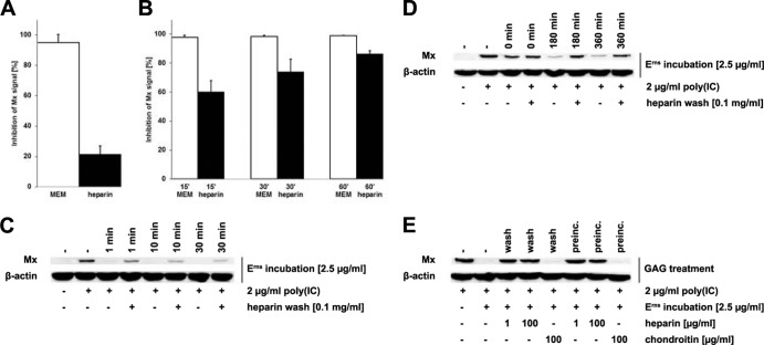
FIG 3.
Erns bound to the cell membrane can be washed off with soluble heparin. Erns was preincubated in wash medium (0.1 mg/ml heparin in MEM) for 30 min before incubation on BT cells for 30 min at 37°C (A). Alternatively, Erns was incubated with BT cells for 15 to 60 min (B) or 1 to 30 min (C), as indicated in the figure, in the absence of heparin prior to treatment with wash medium. Thereafter, BT cells were stimulated with 2 μg/ml poly(I·C) for 18 h at 37°C and cytosolic extracts were assayed as described in the legend to Fig. 1. The signal intensities of Mx synthesis were quantified relative to the levels of β-actin expression (A, B), with complete inhibition of Mx expression being set equal to 100% (mean ± SD, n = 3). Similarly, Erns was incubated for up to 6 h prior to washing, as described above for panels B and C, but at 4°C instead of 37°C (D). To compare the inhibitory activity of heparin and chondroitin, Erns was either preincubated at 4°C in the presence or absence of glycosaminoglycans (preinc.) or incubated on BT cells for 15 min at 4°C prior to washing with either heparin or chondroitin sulfate, followed by Western blotting as described in the legend to Fig. 1 (E). Typical results out of two (C) or three (D, E) independent experiments are shown.

