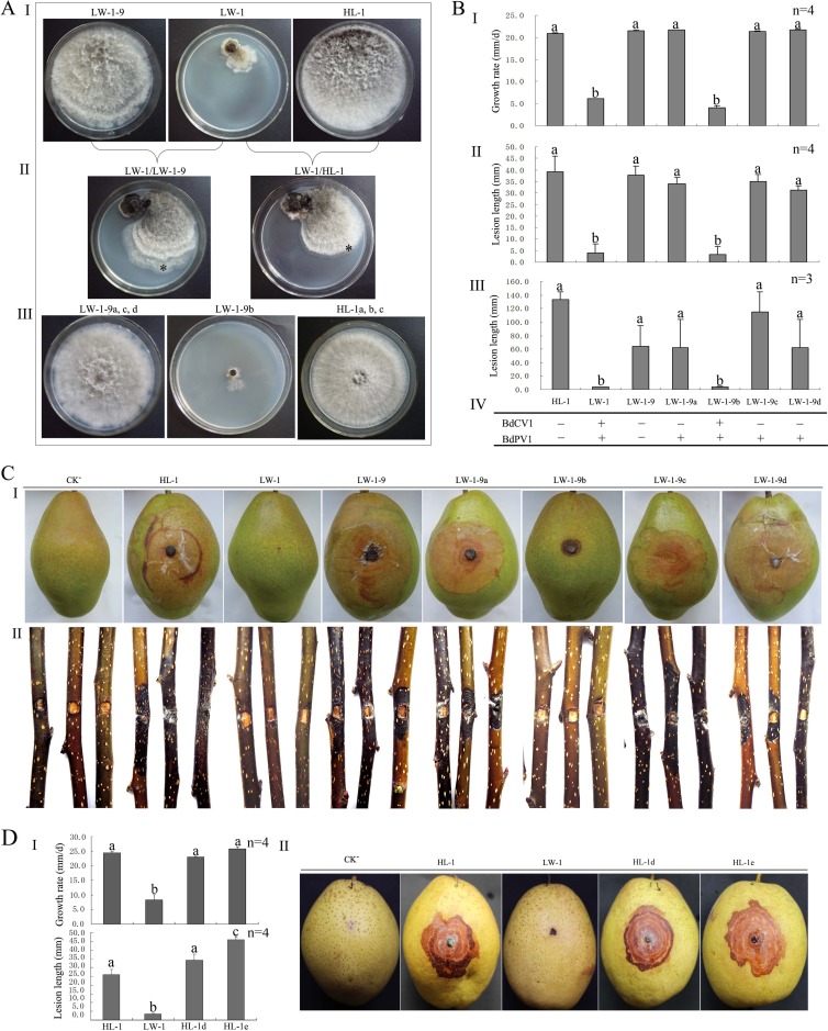
FIG 5.
Horizontal transmission of BdCV1 and BdPV1, growth rate on PDA, and fruit lesion length and tests of virulence of different strains of B. dothidea on pear. (A) Colony morphology of LW-1, LW-1-9, and HL-1 in single culture (I) and contact culture (II) and of subisolates derived from the colony margins of the recipient strains (III). *, location from which a mycelial agar plug was removed for the generation of a derivative isolate for HL-1 or LW-1-9. (B) Histograms of growth rates of subisolates derived from the contact cultures and the parent strains (I), of the length of lesions induced on fruits (II) and branches (III) of pear (P. pyrifolia nakai cv. ‘Hongxiangsu'), and of the presence of BdPV1 and BdCV1 (IV). + and −, the presence and absence of BdPV1 or BdCV1, respectively, on the basis of the results of dsRNA detection by 1.2% agarose gel electrophoresis. (C) Virulence of HL-1, LW-1, and subisolates on fruits (I) and branches (II) of pear (P. pyrifolia nakai cv. ‘Hongxiangsu'). (D) Histograms of the growth rates of subisolates derived from protoplast transfection and the parent strains (I) and of the length of lesions (II) shown by virulence tests (III) on fruits of pear (P. bretschneideri Rehd. cv. ‘Mili'). CK−, treatments inoculated with noncolonized PDA plugs. Bars in each histogram labeled with the same letters are not significantly different (P > 0.05) according to the least-significant difference test.

