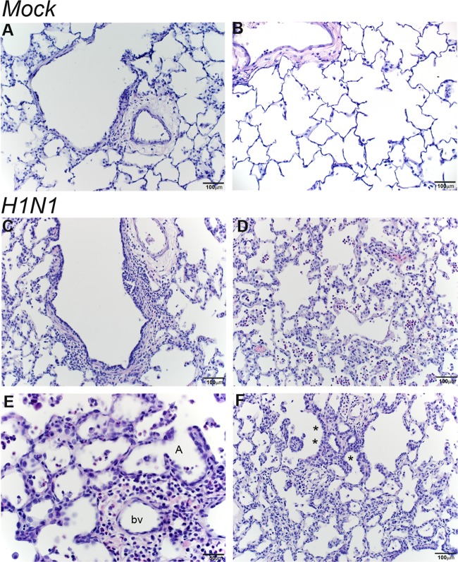
FIG 11.
Persistent inflammation in respiratory bronchioles and alveoli at day 14 post-H1N1 infection. Histological sections from the lungs of mock- and H1N1-infected infant monkeys at 14 days postinoculation were stained with H&E. (A and B) Normal terminal bronchiole (A) and normal alveolar parenchyma (B) from mock-infected animals. (C to E) H1N1-infected animals show bronchiolar walls expanded by inflammation (macrophages and small lymphocytes, with fewer neutrophils) (C) and alveolar inflammation (predominantly neutrophils and macrophages) with thickened alveolar septae (D and E). (F) Alveolar septal thickening with some type II pneumocyte hyperplasia, marked with an asterisk, was also observed for H1N1-infected animals. bv, blood vessel; A, alveoli.

