FIG 3.

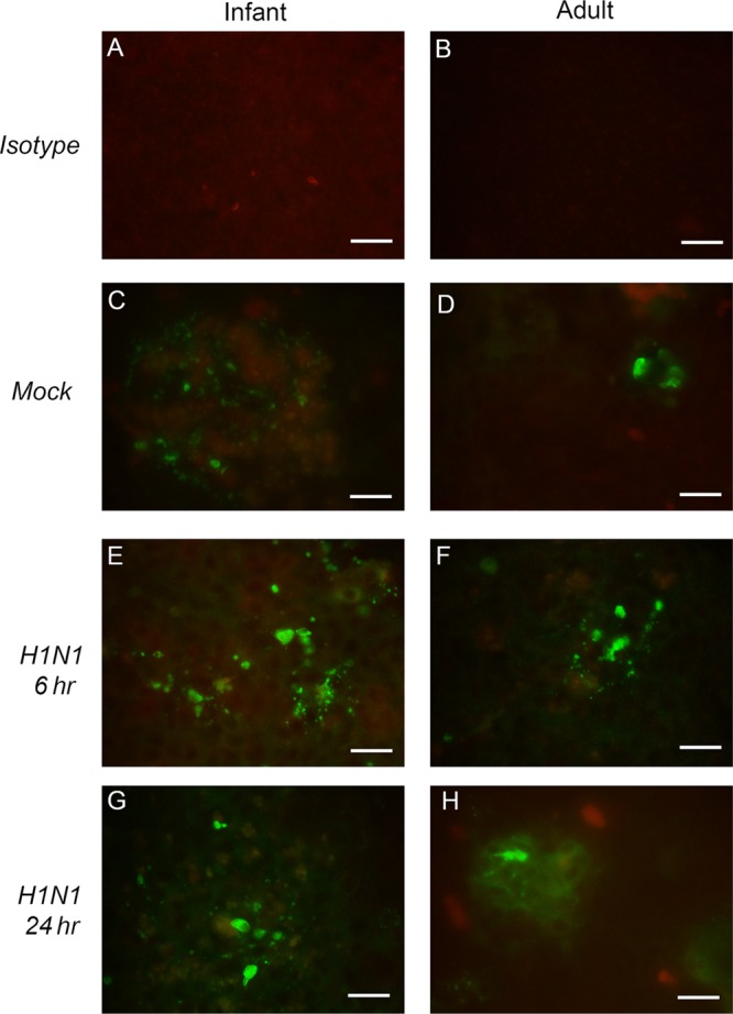
Cleaved-caspase-3 immunofluorescent staining in infant and adult epithelial cell cultures. Representative mock- and H1N1-infected (MOI of 1) epithelial cell cultures were stained with mouse monoclonal antibody to cleaved caspase-3 and visualized with an Alexa 488 fluorescent secondary antibody to detect apoptotic cells. (A and B) Isotype controls are included for comparison. (C to H) Mock-infected cultures were evaluated at 24 h postinfection (C and D), and H1N1-infected cultures were evaluated at 6 h (E and F) and 24 h (G and H) postinfection. Images were collected at a ×20 magnification. Bar = 100 mm.
