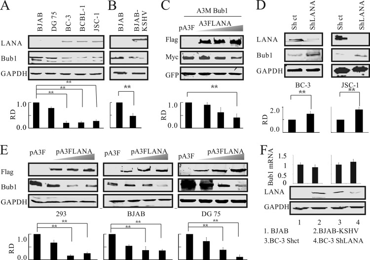
FIG 1.
Bub1 levels are downregulated in cells latently infected with KSHV and expressing LANA. (A) Western blotting was used to detect the endogenous protein levels of Bub1 in KSHV-negative (BJAB and DG75) and KSHV-positive (BC-3, BCBL-1, JSC-1) cell lines. (B) Level of Bub1 protein in BJAB cells infected with KSHV. (C) LANA decreases the level of exogenous Bub1 protein in HEK-293 cells. HEK-293 cells were electroporated with increasing amounts of Flag-tagged LANA, Myc-tagged Bub1, and a GFP plasmid. Forty-eight hours later, the cells were collected for Western blot analysis, which was performed using the indicated antibodies. GFP served as a control for protein loading. (D) LANA knockdown increases Bub1 accumulation. Cell lysates from KSHV-positive B cells (BC-3 and JSC-1) in which LANA or a luciferase control had been stably knocked down (ShLANA or Shct, respectively) were subjected to Western blot analysis. (E) LANA decreases the level of endogenous Bub1 protein in HEK-293 cells and KSHV-negative B cells. HEK-293 and KSHV-negative B cells (BJAB, DG75) were electroporated with increasing amounts of Flag-tagged LANA. Forty-eight hours after electroporation, the cells were collected for Western blot analysis. (F) KSHV infection or LANA overexpression does not affect the mRNA level of Bub1. Total RNAs were isolated from KSHV-infected B cells (BJAB-KSHV, BJAB) and from control knockdown and LANA knockdown BC-3 cells (BC-3 Shct and BC-3 ShLANA, respectively), and the mRNA levels of Bub1 were analyzed by real-time PCR. The relative densities (RD) of Bub1 were quantified and plotted against the signal obtained from the control after normalization to glyceraldehyde-3-phosphate dehydrogenase (GAPDH) (for endogenous Bub1) or ectopically expressed GFP (for exogenous Bub1). Statistical significance was evaluated by using P values of <0.05 (*) and <0.01 (**).

