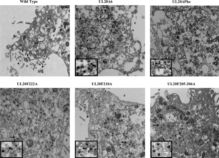
FIG 6.
Ultrastructural morphologies of parental wild-type and mutant viruses. Electron micrographs of Vero cells infected with different viruses at an MOI/cell of 3 and processed for electron microscopy at 18 h.p.i. are shown. The extracellular space (e) and cytoplasm (c) are marked. Arrows show viral particles with envelopment defect, magnified in insets.

