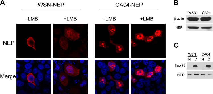
FIG 1.
The cellular localization pattern of CA04-NEP is different from that of WSN-NEP when overexpressed in cells. (A) N-terminally c-Myc-tagged NEP from WSN or CA04 was transiently expressed in 293T cells. At 24 h posttransfection, the cells were treated with 100 ng/μl of cycloheximide for 3 h to block protein synthesis. LMB (11 nM) was added to the medium along with the cycloheximide for LMB treatment. (B and C) Western blotting confirmed the different cellular localizations of the two NEPs. 293T cells transfected with WSN-NEP or CA04-NEP were lysed with whole-cell lysis buffer (B) or nucleus (“N”)-cytoplasm (“C”) fractionation buffer (C) at 24 h posttransfection and prepared for Western blotting. An anti-c-Myc monoclonal antibody was used for protein detection. DAPI (4′-6-diamidino-2-phenylindole) was used for nuclear staining. Results shown are representative images.

