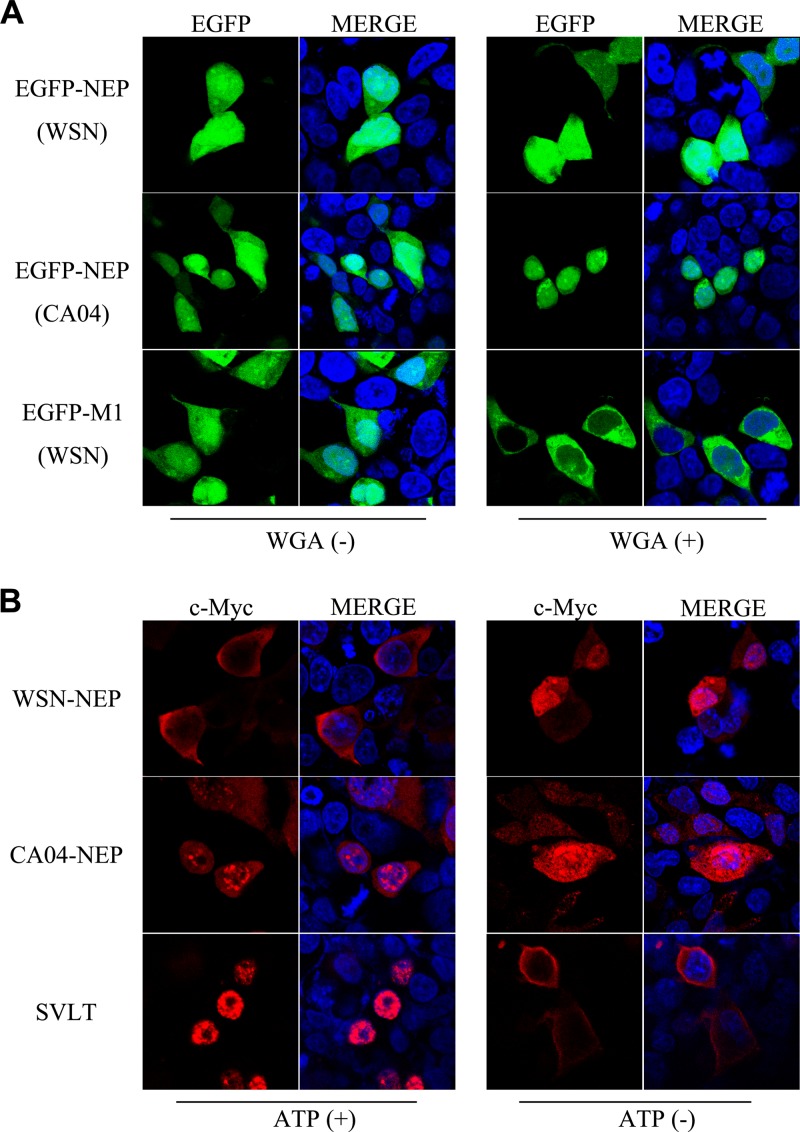
FIG 3.
NEP enters the nucleus by passive diffusion. (A) In vitro NEP transport assay. EGFP-NEP (upper panel) and EGFP-M1 (lower panel) fusion proteins were separately expressed in 293T cells, and cell lysates in transport buffer were added to digitonin-treated 293T cells. For WGA treatment (WGA+), the permeabilized cells were incubated in transport buffer containing 50 μg/ml WGA before addition of cell lysates. Images revealed fluorescence signals of EGFP from fusion proteins. (B) Energy depletion assay of the nucleocytoplasmic transport of NEP. N-terminally myc-tagged NEP was expressed in 293T cells. N-terminally FLAG-tagged SVLT was expressed as a positive control. At 24 h posttransfection, the cells were incubated with energy depletion medium (ATP−) or normal culture medium (ATP+) for 3 h before fixation and visualization. Anti-myc and anti-FLAG monoclonal antibodies were used for the detection of the indicated proteins.

