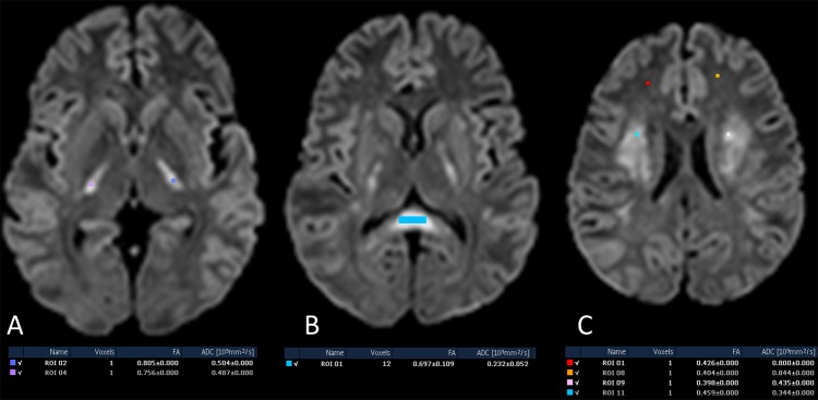
Figure 3.

At admission: axial diffusion-weighted images with estimation of fractional anisotropy (FA) values; at splenium of corpus callosum (A), posterior limbs of internal capsule (B) and centrum semiovale (C) showing normal or mildly increased FA values with reduced apparent diffusion coefficient (ADC) values as compared to normal-appearing white matter in bilateral frontal lobes (C).
