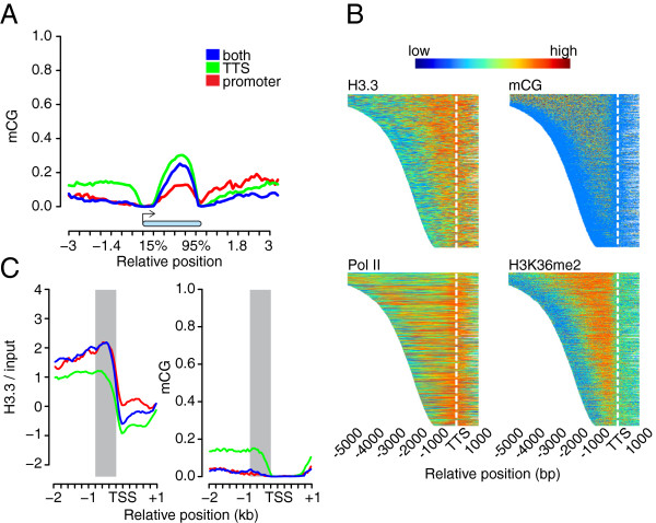
Figure 6.

DNA mCG methylation at promoters does not coincide with H3.3 enrichment. (A) Metagene plot of mCG across gene bodies (blue bar) between -3 kb and +3 kb. mCG data are from [40]. Genes were grouped according to the presence of H3.3-containing nucleosomes in the promoter (red), close to the TTS (green) or both (blue). (B) H3.3, mCG, Pol II and H3K36me2 profiles around TTS. Signals were plotted from the TSS up to 1 kb proximal to the TTS for 5,000 random sampled genes ordered by their gene body lengths. For genes that are longer than 5 kb, only 5 kb of the gene body are shown. Epigenome profiles are represented by a heat map color code with red and blue representing highest and lowest values, respectively. White dashed lines indicate the TTS positions. Data are from [23,29,40]. (C) Metagene plot of H3.3 signal (left) and mCG (right) between -2 kb and +1 kb of the TSS. mCG data are from [40]. Genes were grouped according to the presence of H3.3-containing nucleosomes at the promoter (red), close to the TTS (green) or both (blue). Grey areas indicate -800 to -200 bp of the TSS.
