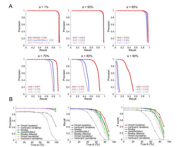
Figure 4.

Performance of somatic mutation detection. Performance comparison of different methods for somatic mutation detection. (A) Precision-recall curves for Virmid with α (red), Virmid without α (light red) and JointSNVMix2 (blue) for six different α values (1%, 50%, 60%, 70%, 80% and 90%). Note that the performance is significantly improved when α is incorporated into the calling model. There is little difference in performance at low contamination levels (α ≤ 50). (B) Precision and recall scores of the final call generated for each α where mutation probabilities are not available; note that a single point instead of a curve is plotted for each α. As α increases, there is a consistent drop in precision, recall and F-score. The latter is given by: Four tools including Virmid, Strelka, VarScan2 and JointSNVMix2 were tested with the same data. Virmid and VarScan2 were tested in two different modes (with and without α). Strelka was also tested in two modes with or without applying quality control. Overall, Virmid with α had the best F-score, followed by Strelka, VarScan2 with α and JointSNVMix2. Note that the tools with α (Virmid with α, Strelka and VarScan2 with α) outperformed those without α (Virmid without α and VarScan2 without α), showing the importance of incorporating α in SNV calling. SNV, single nucleotide variation.
