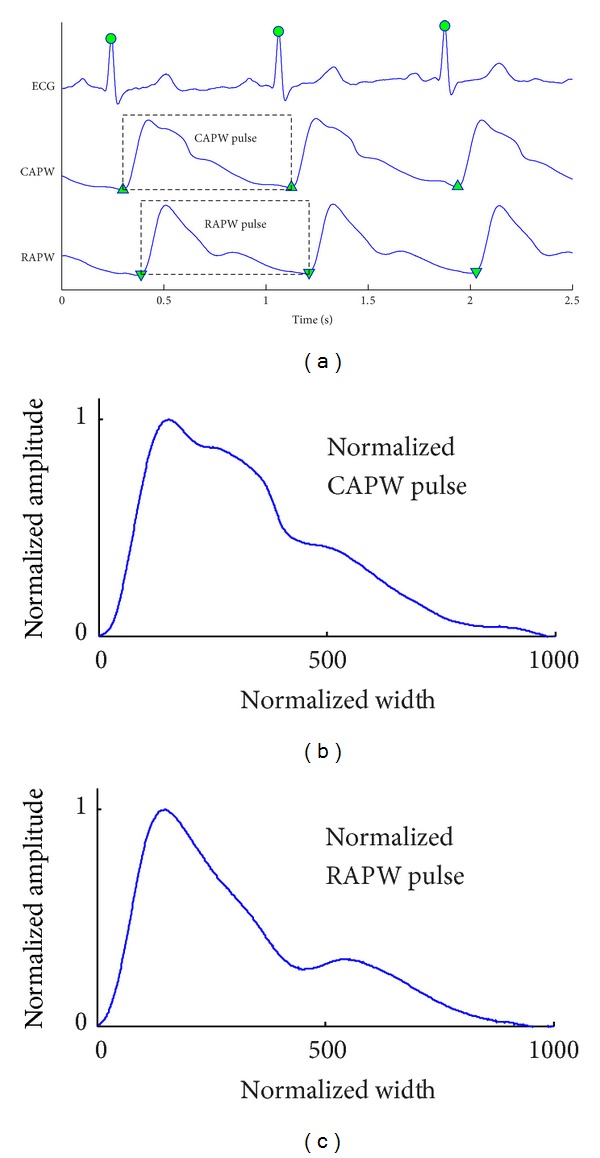
Figure 1.

(a) One example of recorded ECG, carotid artery pressure waveform (CAPW), and radial artery pressure waveform (RAPW) signals. The detected R-wave peaks are denoted by “●”, and the starting points of CAPW and RAPW signals are denoted by “▲” and “▼,” respectively. (b) Normalized CAPW and (c) RAPW pulses with width up to 1000 points and the amplitude to unity between 0 and 1.
