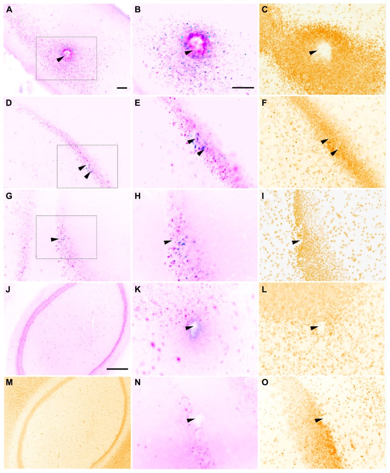
FIGURE 3.
Representative horizontal sections through the dorsal hippocampus of rats implanted with a macroelectrode (A–C), a microelectrode array (D–F), or a sonicoplated microelectrode array (G–I) stimulated and then stained for c-fos (blue), NeuN (pink) and the nuclear marker DAPI (mustard). Macroelectrode and microelectrode implanted dorsal hippocampal sections from rats that did not receive any stimulation are shown in K–L and N–O respectively. J,M are sections of the dorsal hippocampus contralateral to the site of macrostimulation. The region within the dotted box in A,D, and G is shown at higher magnification in B,E, and H, and C,F, and I. Location of implanted electrodes is indicated with arrows. Scale bar (for all except J,M): 100 μm. J,M Scale bar: 500 μm.

