Figure 3.

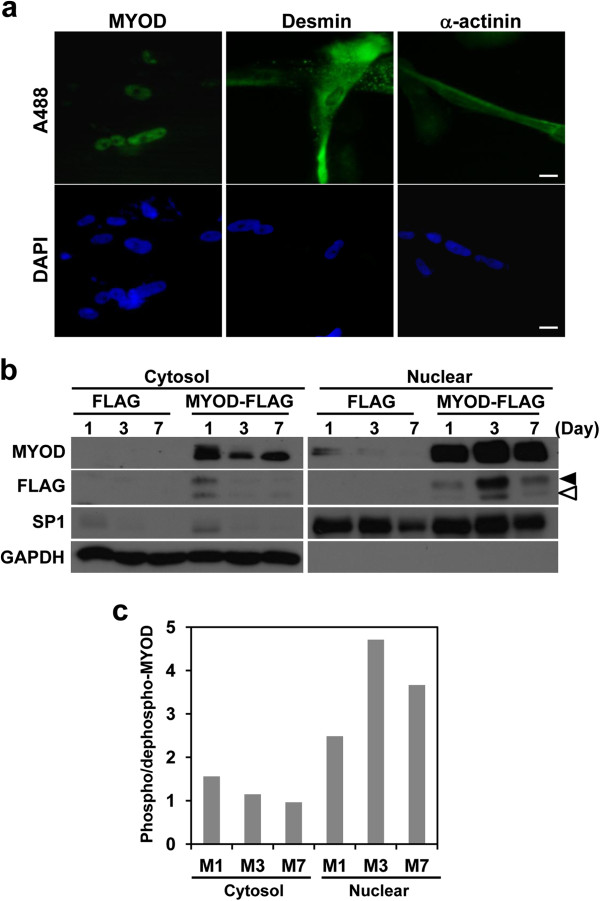
Intracellular localization of MYOD, desmin and α-actinin proteins during myogenesis. (a) Immunocytochemistry of hAFS cells infected with MYOD lentivirus. The infected cells were cultured in myogenic differentiation medium for seven days. Myogenic marker proteins, MYOD, desmin and α-actinin were immunostained with monoclonal antibodies and Alexa Fluor 488 conjugated secondary antibody (upper). Nuclei were stained with DAPI (lower). (b) Western blotting of MYOD protein in the nuclear-cytosol fractions of differentiating hAFS cells at 1, 3 and 7 days. hAFS cells were transduced with FLAG pHJ-1 (FLAG) or MYOD-FLAG pHJ-1 (MYOD-FLAG). Phosphorylated MYOD (black arrow head, 47 KDa) and dephosphorylated MYOD (white arrow head, 45 KDa) are indicated. (c) The ratio of phosphorylated and dephosphorylated MYOD during myogenic differentiation. The band densities of Flag tagged MYOD at (b) were measured and the densities were calculated as phosphorylated/dephosphorylated MYOD (Scale bar = 10 μm). DAPI, 4',6-diamidino-2-phenylindole; hAFS, human amniotic fluid stem.
