
Fig. 7.
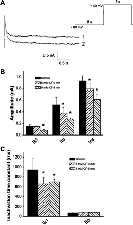
Inhibition of Ik1 by external Cl−. A: superimposed pairs of currents at +40 mV recorded following conditioning pulses (−80 mV) for control 150 mM Cl− (trace 1) and 5 mM Cl− (trace 2). Red solid lines represent exponential fitting. Stimulation protocol is depicted at top right. B: time-dependent inhibition of current amplitude of Ik1, Ito, and steady-state current (Iss). C: time-dependent inhibition of inactivation time constant of Ik1 by external Cl−. *Significance at the 0.05 level.