Figure 6.

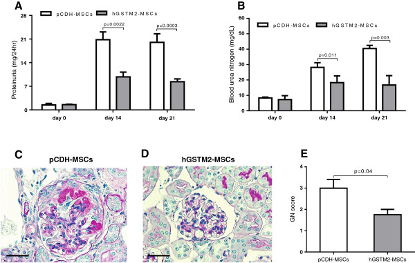
Human glutathione S-transferase Mu 2 mesenchymal stem cells (hGSTM2-MSCs) attenuated anti-glomerular basement membrane antibody-induced glomerulonephritis (anti-GBM-GN) in 129/svj mice. (A-B) Proteinuria and BUN were measured in 24-hour urine samples on days 0, 14, and 21 in anti-GBM-challenged 129/svj mice injected with 1 × 10⁶ pCDH-MSCs (vector controls) or the same number of hGSTM2-MSCs. (C-D) Renal histological images from (C) pCDH-MSC-injected and (D) hGSTM2-MSC-injected 129/svj mice challenged with anti-GBM antibody. Renal sections were stained with periodic acid Schriff (PAS). (C) Representative images of the kidney of pCDH-MSC-injected mice, showing some of the typical light microscopic features seen in anti-GBM-induced nephritis in 129/svj mice, including intracapillary hypercellularity, with obliteration of the capillary lumina, and tubular dilatation with casts. (D) Representative images of the kidneys of the anti-GBM-GN mice injected with hGSTM2-MSCs, showing mild renal histological changes. (E) Renal pathology score of anti-GBM-challenged 129/svj mice treated with pCDH-MSCs and hGSTM2-MSCs. Images are representative of sections from at least five mice in each study group (original magnification ×400, scale bars = 50 μm).
