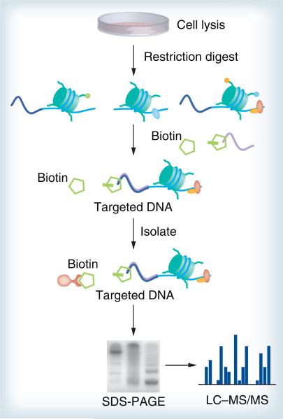
Figure 2. Proteomics of isolated chromatin.
After cell lysis and restriction digestion, the targeted loci are captured by primers that bind to a specific genomic region. An enzymatic step incorporates biotin labels only to chromosomal fragments that contain the targeted sequence and streptavidin-coated magnetic particles isolate the targeted chromatin. Proteins associated with the isolated regions are separated by SDS-PAGE and then analyzed by MS.
LC–MS: Liquid chromotography–mass spectrometry; MS: Mass spectrometry.

