
Figure 8. iBALT formation in wild-type and STAT6-deficient mice upon Ad-mOSM administration.
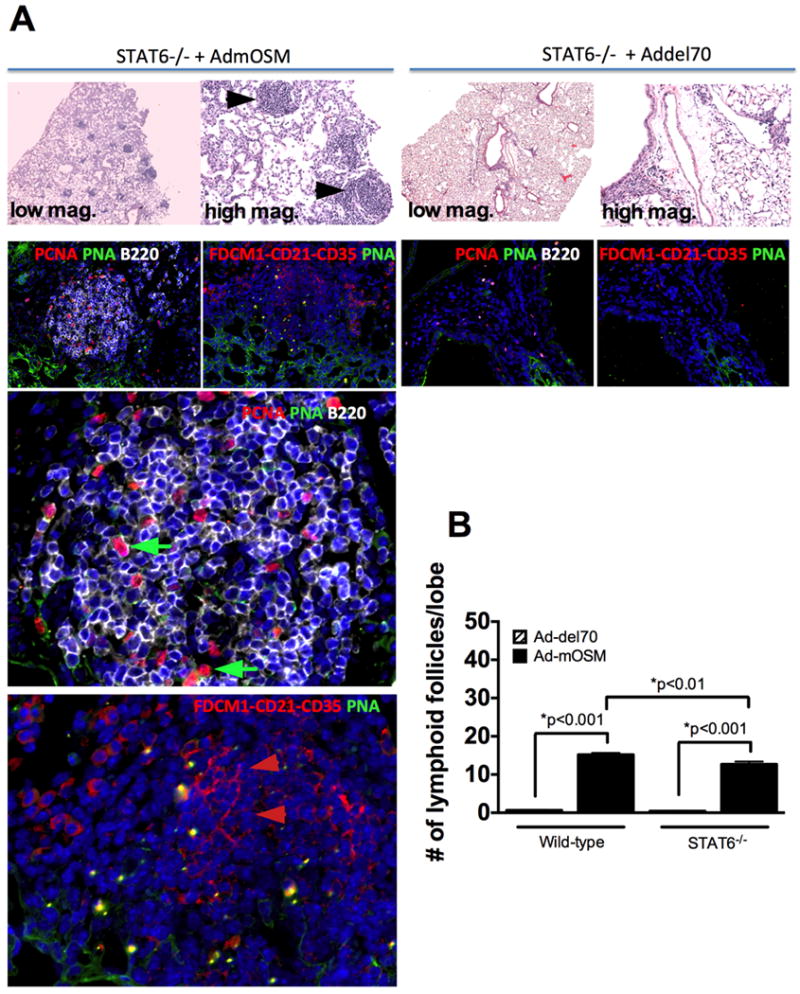
C57Bl/6- STAT6-gene-deficient mice received Ad-del70 or Ad-mOSM, as described in Figure 1. 10% formalin-fixed lung tissues were examined for the presence of lymphocytic aggregates in H&E stained slides (A, top panels at 50× and 200× magnification). Complexity of iBALT structures was evaluated by staining for B220 (B cells, white surface stain) and PCNA (red nuclear stain) in (A) middle panels. Germinal center B cells were detected with peanut agglutinin (PNA, green surface stain, green arrows). Presence of FDC cells in the iBALT structures was assessed with a combination of antibodies against FDC antigen, CD21, CD35 (red, red arrows). The two bottom staining images are magnifications of the 200× images from the Stat6-/- mice treated with Ad-mOSM. (B) Number of lymphoid follicles per lung lobe in C57BL/6- and Stat6-/- mice, infected with Ad-del70 or Ad-mOSM are shown. Results are presented as the means ± SEMs of at least five mice per group. Data shown is representative of at least two separate experiments.