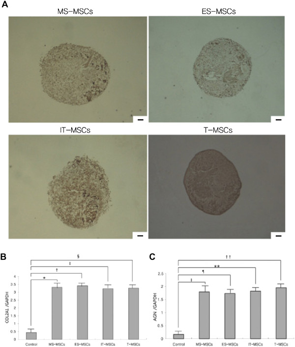
Figure 5.

Comparative analysis of chondrogenic differentiation capacity of MS-, ES, IT- and T-MPCs. (A) Chondrogenesis was demonstrated by the formation of a sphere and type II collagen expression. There were no differences in the size of sphere and the immunohistochemical staining for type II collagen among any of the MPCs groups (original magnification 100×, scale bar = 50 μm). The expression of specific chondrogenic genes, COL2A1(B) and AGN(C), was evaluated by real-time qRT-PCR. COL2A1 and AGN were up-regulated during chondrogenesis in all MPCs groups. However, there were no significant differences in the expression level of two markers among any of the MPCs groups. Data are expressed as the mean ± SEM. *, †, ‡, ǁ, ¶, ** P <0.001, § P = 0.002, †† P = 0.005. AGN, aggrecan; COL2A1, collagen type II α1; ES, ethmoid sinus; IT, inferior turbinate; MPCs, mesenchymal progenitor cells; MS, maxillary sinus; T, tonsil.
