
Figure 5.
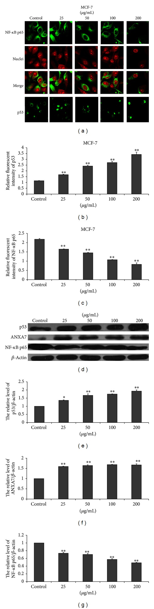
EECP regulated the levels of ANXA7, p53, and NF-κB p65 in MCF-7 cells. (a) Fluorescent micrographs obtained at 48 h (×400). Nuclei were counterstained with PI. (b) and (c) The relative fluorescence intensity of NF-κB p65 and p53 in MCF-7 cells. (d) The levels of p53, ANXA7, and NF-κB p65 were detected by western blot at 48 h. (e), (f), and (g) The hemiquantification of p53, ANXA7, and NF-κB p65 levels in MCF-7 cells (*P < 0.05, **P < 0.01 versus control, n = 3).