
Figure 3.
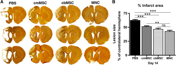
All three cell types promoted histopathologic recovery, with greater improvement seen in the cbMNC group. Silver-stained brain sections showed decreased infarct size, reduced ventricular enlargement, and loss of cytoarchitecture in all three cell-treated animals compared with the PBS group (A). Significant reduction in percentage of infarct area across all three cell groups (P < 0.001) was seen compared with the PBS group at d 14 after stroke (B). Reduction in lesion size was significantly more in the cbMNC versus cmMSC group (P < 0.01). Values are expressed as mean ± SEM. (**) is P < 0.01; (***) is P < 0.001, and (ns) is nonsignificant.