
Figure 2.
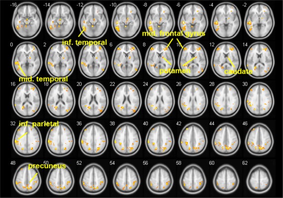
Comparing anterior cingulate cortex (ACC)-seeded resting-state functional connectivity (rsFC) in smokers during the abstinent condition to that of nonsmokers. Smokers showed greater rsFC in the precuneus, caudate, putamen, superior frontal gyrus, middle frontal gyrus, superior parietal gyrus, superior occipital lobe, inferior parietal lobe, middle temporal lobe, and the inferior temporal gyrus (P < 0.05). There were no areas showing decreased rsFC in smokers in the withdrawal condition as compared to controls. Brighter color indicates a higher t value.