
Figure 3.
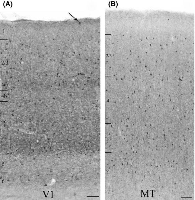
Qualitative comparison of single-label immunoperoxidase reactivity for parvalbumin in V1 (A), and the middle temporal visual area (MT) (B). Parvalbumin (PV) neurons are present in cortical layers 2 through 6 in both areas. In V1 there are occasional PV neurons in layer 1 as well (arrow). There is denser staining of the neuropil and an apparently higher density of somata in the thalamic recipient layers of V1 (layers 4a, 4c, and 6). There is no comparable banding in MT, where the neuropil staining is generally more diffuse. The micrographs present images of different, but coprocessed tissue sections from a single animal. Layer boundaries are indicated on the left of each panel. Imaged with a 10× objective. Scale bars = 100 μm.