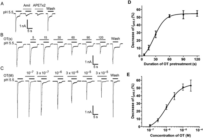
Figure 1.
Inhibition of ASIC currents by OT in rat DRG neurons. (A) Representative current traces were evoked by extracellular application of a pH 5.5 solution for 5 s in the presence of capsazepine (10 μM) to block proton-induced TRPV1 activation. The proton-induced current could be completely blocked by 100 μM amiloride (Amil), a broad-spectrum ASIC channel blocker. It was also blocked by 3 μM APETx2, an ASIC3 blocker. (B and D) Representative current traces and summary data show the effect of OT pretreatment duration on IpH. Current traces illustrate the effect of OT (10−5 M) pretreatment duration on IpH in a DRG cell. The graph shows that the inhibitory effect of OT (10−5 M) increased as OT pretreatment duration increased from 15 to 120 s (n = 8). (C and E) Representative current traces and summary data show a concentration-dependent inhibition of the peak amplitude of IpH by OT. The sequential current traces illustrate the inhibition of IpH by different concentrations of OT (10−7 M − 3 × 10⁻⁵ M). All data were obtained from a single DRG neuron. The graph shows OT decreased IpH in a concentration-dependent manner. Each point represents the mean ± SEM of seven to nine neurons. DRG neurons with membrane potential clamped at −60 mV.

