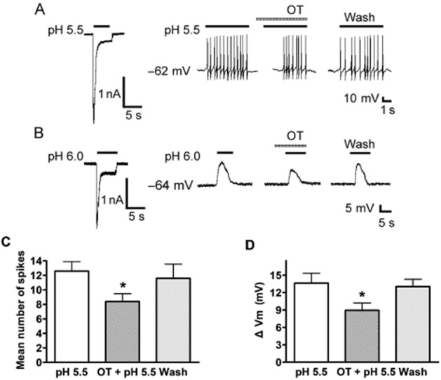
Figure 4.

Effect of OT on proton-evoked membrane excitability of rat DRG neurons. (A) Original current and spikes recordings from the same DRG neuron. Left panel: a pH 5.5 acid stimulus induced an inward current with voltage-clamp recording. Right panel: the pH 5.5 acid stimulus produced cell spikes with current-clamp recording in the same neuron in the presence of the TRPV1 inhibitor capsazepine (10 μM). The treatment with OT (10−5 M) for 60 s inhibited the acidosis-induced spiking activity. (B) Original current and membrane potential recordings from the same DRG neuron. Left panel: voltage-clamp recording of current induced by a pH 5.5 acid stimulus. Holding potential was −60 mV. Right panel: current-clamp recording (I = 0 pA) of the depolarization evoked by the pH 5.5 acid stimulus from the same neuron as the left panel. The treatment of OT (10−5 M) for 60 s decreased the acidosis-induced membrane depolarization. No action potential was triggered by the membrane depolarization in the neuron tested in the presence of capsazepine (10 μM) and TTX (1 μM) to block proton-induced TRPV1 activation and Na+ channel-mediated action potentials respectively. (C and D) Bar graphs show the effects of OT on the number of spikes and membrane potential depolarization produced by pH 5.5. The acidosis-evoked depolarization and spikes recovered to control condition after washout of OT for 10 min. *P < 0.05, paired t-test, compared with pH alone, n = 10 in each column.
