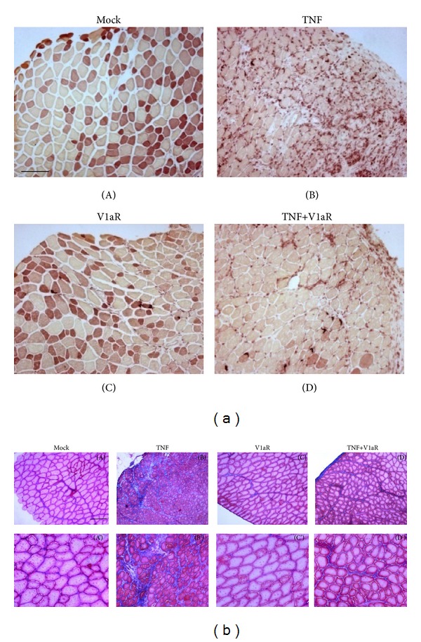
Figure 3.

V1aR overexpression attenuates inflammation and fibrosis induced by high levels of TNF. (a) Nonspecific esterase staining of TA cross-sections, performed one week after electroporation. The figure highlights the massive presence of macrophages in muscle overexpressing TNF (B) and a reduced esterase activity in muscle overexpressing both TNF and V1aR (D). (Scale bar = 50 μm, magnification 10x.) (b) Masson's trichrome stain of TA cross-sections, one week after electroporation, demonstrates less extensive fibrosis in muscle overexpressing TNF+V1aR (D and D'), compared with muscle overexpressing TNF alone (B and B'). (Scale bar = 50 μm, magnification 10x, (A)–(D); 20x, (A')–(D').
