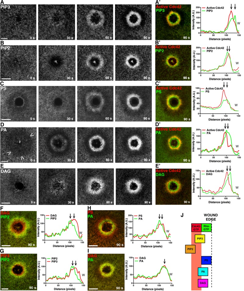
FIGURE 1:
Multiple, distinct lipid domains form around single-cell wounds. (A) eGFP-PH (GRP1) detects PIP3 accumulation. (A′) PIP3 peak enrichment occurs inside the active Cdc42 zone at 90 s. (B) eGFP-PH (PLCδ) detects PIP2 enrichment. (B′) PIP2 peak enrichment is outside the active Cdc42 zone at 90 s. (C) eGFP-C2 (lactadherin) detects PS enrichment. (C′) PS peak enrichment occurs inside the active Cdc42 zone at 90 s. (D) eGFP-Spo20 detects a region of PA depletion (arrowheads) immediately after wounding, followed by enrichment. (D′) The peak of PA signal is found inside the zone of active Cdc42 at 90 s. (E) eGFP-C1 (PKCη) detects a region of DAG enrichment. (E′) The peak of DAG enrichment leads the peak of active Cdc42 at 90 s. (F) The peak of DAG enrichment leads the peak of PIP2. (G) The peak of PIP3 enrichment leads the peak of PIP2. (H) The peak PS signal leads the peak PA signal. (I) The peak DAG and PA signal are colocalized. (J) Schematic of lipid localization relative to the active Rho and Cdc42 zones. Scale bar, 20 μm. W, wound.

