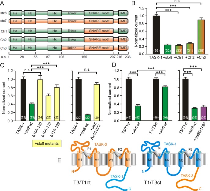
FIGURE 3:
Dissection of the interacting regions of stx8 and TASK-1. (A) Topology of stx8, stx7, and the stx8/stx7 chimeras. (B) TASK-1 currents measured in Xenopus oocytes expressing TASK-1 and stx8 or stx8/stx7 chimeras. (C) Normalized hTASK-1 currents measured in Xenopus oocytes expressing hTASK-1 and stx8 or deletion mutants of stx8. (D) Normalized currents measured in Xenopus oocytes expressing TASK-3/TASK-1 or TASK-1/TASK-3 chimeras alone or together with stx8 or stx8Q179A. (E) Schematic drawing of TASK-3/TASK-1 and TASK-1/TASK-3 chimeras.

